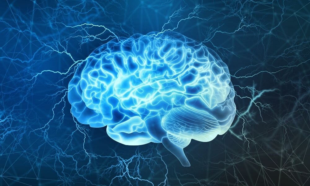
All memory storage devices store information by modifying their physical properties from your brain to your computer’s RAM. Cajal recommended that the brain stores a lot of information’s by rearrange relations, or synapses, between neurons.
Since then, neuroscientists have sought to understand the physiological changes associated with memory formation. But synapses are hard to imagine and map. They are a million times smaller than the minor thing a standard medical MRI could imagine. In addition, the brains of mice contain about 1 billion synapses, which researchers often use to study brain functions, and they are as vague to all translucent colors as the tissues around them.
However, a fresh imaging technique developed by my mates and me has permissible us to map synapses throughout memory configuration. We noticed that new memories depend on how cells make connections with each other. While some parts of the brain produce more connections, others lose them.
Mapping new memories in fish
In their research papers, researchers in the past were focusing and recording only on electrical signals generated by neurons. Although these experiments have confirmed that neurons change their response to specific stimuli after the memory is formed, they have not pinpointed what triggered these changes.
To see how the brain physically changes when the brain builds new memory, we created 3D maps of zebrafish synapses of memory formation before and after. We chose zebrafish as our first test subjects because their brains are large enough to function just like humans but are small and transparent enough to present a window into the living brain.
To create a new memory in fish, we used a learning method called classical conditioning. Classical conditioning includes exposing an animal to two different stimuli simultaneously: a neutral one that does not give a reaction and an unpleasant one that the animal tries to avoid. When these two stimuli are combined several times, the animal responds to the neutral stimulus as if it were an unpleasant stimulus, indicating that it binds these stimuli together to form an associative memory. Has taken
We lightly warmed the fish’s head with an infrared laser as an unpleasant stimulus. When the fish wagged its tail, we took it as a signal that it wanted to leave. When the fish is first exposed to a neutral stimulus, the light is turned on, and the tail jerk means what happened when it first encountered the unpleasant stimulus.
We genetically engineered zebrafish with neurons that produce more fluorescent proteins that attach to synapses and make them visible to create the maps. We then photographed Synapses with a custom-made microscope, which used a much lower dose of laser light than standard instruments, and also used fluorescence to create images. Because our microscopes did little damage to neurons, we were able to capture images of synapses without losing their structure and function.
After Mapping 3D synapse maps before and after memory formation, we came to know that neurons in an area of the brain, the anterolateral dorsal pallium, developed new synapses, while neurons mainly in other regions. Anteromedial dorsal pallium lost synapses. This meant that new neurons connected while others destroyed their connections. Experiments in the past have suggested that the fish’s dorsal palm may resemble the amygdala of mammals, where memories of fear are preserved.
Surprisingly, the changes in the power of existing connections between neurons that occurred with memory formation were small and not different from the changes in control fish that did not create new memories. This means that the formation of an associative memory involves the formation and loss of synapses but does not necessarily change the strength of existing synapses, as previously thought.
Could removing synapses remove memories?
Our new way of observing the work of brain cells can open the door to an in-depth understanding of how memory works and possible ways to treat neuropsychiatric conditions such as PTSD and addiction.
Associative memories
Associative memories are much stronger than other types of memories, such as conscious memories of what you ate for lunch yesterday. Moreover, memories stimulated by classical conditioning are thought to resemble traumatic memories that cause PTSD. Otherwise, harmless stimuli, such as those experienced by someone during a trauma, can cause painful memories to be recalled. For example, bright light or a loud voice can bring back memories of a battle. Our study shows the role those synaptic connections can play in memory and could explain why peer-to-peer memories can last longer and be remembered more clearly than other types of brain memories.
Currently, the most common treatment for Post-Traumatic Stress Disorder PTSD, exposure therapy, involves repeatedly exposing the patient to a harmless but dynamic stimulus to suppress the memory of the traumatic event. In theory, it indirectly regenerates brain synapses to make memory less painful. Although exposure therapy has had some success, patients are at risk of recurrence. This indicates that the underlying memory that caused the painful reaction has not been eliminated. Conceptually linked to classic conditioning, long exposure therapy is one way to treat PTSD.
It is unknown whether synapse generation and damage trigger memory formation. My laboratory has developed a technology t can quickly, precisely and accurately remove synapses without damaging neurons cells. We plan to use similar methods and ways to remove synapses in zebrafish or mice to see if they alter harmonious memories.
In these ways, it may be possible to physically erase associative memories of enduring devastating conditions such as PTSD and addiction. However, before such a treatment can be considered, the synaptic changes encoding memory memories need to be described more clearly. Some serious issues come in ethical and technical barriers that must be included.

