
The lens shows a wide view, but still offers a sense of human-like depth perception: as close objects come into focus, far away objects look blurry.
“Our eye can change focus. An insect eye is made of many small optical components that can’t change focus but give a wide view. We can combine the two,” said Yi Zhao, associate professor of biomedical engineering and ophthalmology at Ohio State University.
“What we get is a wide-angle lens with depth of field,” said Zhao.
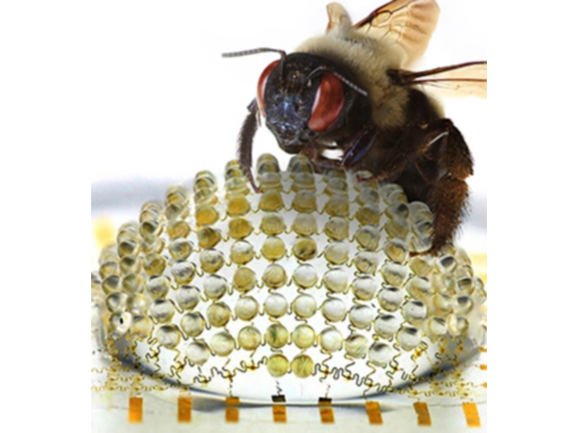
The prototype lens is made of a flexible transparent polymer filled with a gelatinous fluid similar to fluid inside the human eye. It’s a composite of several separate dome-shaped fluid pockets, with small domes sitting atop one larger dome.
Each dome is adjustable, so that as fluid is pumped into and out of the lens, different parts of it expand and contract to change the overall shape?and thus, the direction and focus?of the lens.
This shape-changing strategy is somewhat similar to the way muscles in the human eye change the shape of the lens tissue in order to focus.
It differs dramatically from the way typical cameras and microscopes focus, which involves moving separate glass lenses back and forth along the line of sight.
The shape-changing lens could potentially offer the same focusing capability as multiple moving lenses in a single stationary lens, which would make for smaller and lighter cameras and microscopes.
Zhao is interested in using the lens in confocal microscopes, which use a system of moving glass lenses and a laser to scan three-dimensional images of tiny objects.
“We believe that it is possible to make a confocal microscope with no moving parts,” he said.
Zhao and doctoral student Kang Wei demonstrated that the lens was able to switch its focus among microscopic objects arranged at different distances.
With laparoscopy, doctors insert tiny wide-angle cameras into the patient’s body in order to see as much tissue as they can without cutting the patient open. But such lenses don’t offer a sense of depth: they show all objects?both near and far?in focus at all times.
This poses a problem for doctors; if they mistake a close object for a far away one, they could accidentally graze healthy tissue with the scope or surgical instruments.
“With our lens, doctors could get the wide-angle view they need, and still be able to judge the distance between the lens and tissue. They could place instruments with more confidence, and remove a tumour more easily, for example,” Zhao said.